Nikon Small World 2021 Photo Competition winners announced
From neurons to tick heads to louse claws, here are the top 10 images from the competition.

The winning image of the Nikon Small World 2021 Photo Competition shows a southern live oak leaf’s trichomes, stomata, and vessels. Image Credit: Jason Kirk, Baylor College of Medicine
The winners of the Nikon Small World Photomicrography Competition, which aims to showcase “the beauty and complexity of life as seen through the light microscope,” were announced today.
This is the 47th year of the photo competition, which is open to anyone with an interest in microscopy—the use of microscopes to view samples and objects—and photography. This year the contest received almost 1,900 entries from 88 countries. A panel of five judges* evaluated the entries for originality, informational content, technical proficiency, and visual impact, Nikon reported. The results of the sister video competition, Small World In Motion, were announced last month.
Here are this year’s top 10 images:
1. Live oak leaf
Related
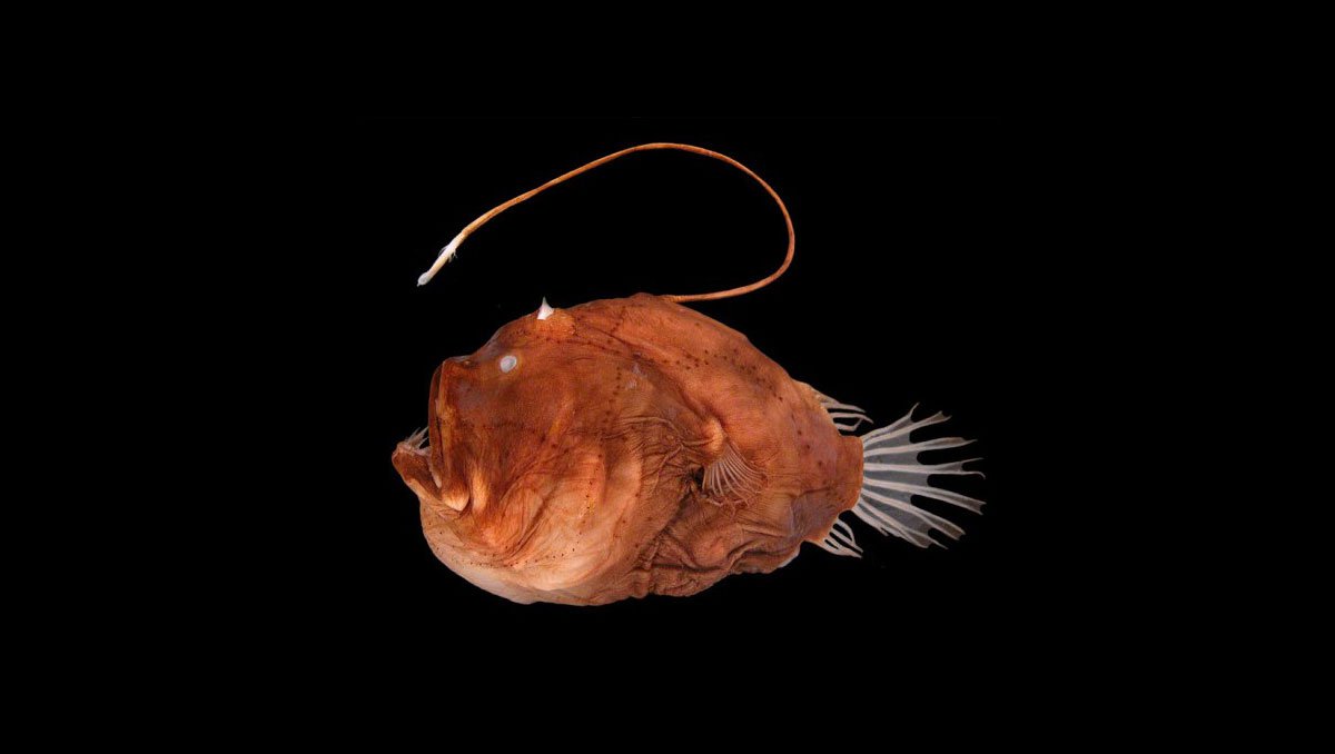
Bioluminescence in fish evolved 27 different times

Creatures of Light

Astronomers Photograph Two Newborn Planets for the First Time
A southern live oak leaf’s trichomes, stomata, and vessels photographed by Jason Kirk, a professional imager and core director of Baylor College of Medicine’s Optical Imaging & Vital Microscopy Core. Kirk used a custom-made microscope system to take around 200 individual images of the leaf, which he stacked together to create this image, Nikon reports.
Trichomes, stomata, and vessels are all “essential to plant life,” Nikon writes. Trichomes, fine outgrowths that protect a plant from extreme weather and insects, are featured in white. “In purple, Jason highlights the stomata, small pores that regulate the flow of gases in a plant. Colored in cyan are the vessels that transport water throughout the leaf,” Nikon said in a statement.
2. Networking neurons in microfluidic device

Image Credit: Esmeralda Paric and Holly Stefen, Dementia Research Centre, Macquarie University, New South Wales, Australia
This image is of a microfluidic device, which contains 300,000 networking neurons divided into two isolated populations (left and right) bridged by axons (center). The isolated populations were each treated with a unique virus, Nikon reports. The image was taken at 40X magnification by Esmeralda Paric and Holly Stefen of Macquarie University’s Dementia Research Centre in New South Wales, Australia, using fluorescence imaging, which uses high-intensity illumination to excite fluorescent molecules in a sample. “When a molecule absorbs photons, electrons are excited to a higher energy level,” Nikon writes. “As electrons ‘relax’ back to the ground-state, vibrational energy is lost and, as a result, the emission spectrum is shifted to longer wavelengths.”
3. Rear leg, claw and respiratory trachea of a louse

Image Credit: Frank Reiser, Nassau Community College, New York
A rear leg, claw, and respiratory trachea of a louse, a wingless parasitic insect, at 5X magnification taken by Frank Reiser, a biologist at Nassau Community College in New York. Reiser used darkfield micrography, which “creates contrast in transparent unstained specimens” and “depends on controlling specimen illumination so that central light which normally passes through and around the specimen is blocked,” Nikon writes, and image stacking to produce this image.
4. Embryonic rat neuron

Image Credit: Paula Diaz, Pontificia Universidad Católica de Chile, Santiago, Chile
A sensory neuron from an embryonic rat taken by Paula Diaz, a physiologist at Pontificia Universidad Católica de Chile in Santiago, Chile. Diaz took the image at 10X magnification and used fluorescence imaging to produce it.
5. Housefly proboscis

Image Credit: Oliver Dum, Medienbunker Produktion, Bendof, Rheinland Pfalz, Germany
A proboscis of a housefly (Musca domestica) taken by Oliver Dum of Medienbunker Produktion in Bendof, Rheinland Pfalz, Germany. Dum took the image at 40X magnification and assembled it using image stacking.
6. Mouse brain vasculature

Image Credit: Dr. Andrea Tedeschi, Wexner Medical Center, Ohio State University
3D vasculature of an adult mouse brain taken by Dr. Andrea Tedeschi of Ohio State University’s Wexner Medical Center in Columbus, Ohio. Tedeschi took the image at 10X magnification and used confocal imaging, which involves scanning a specimen to create extremely thin (down to 250 nanometer thickness) computer-generated optical sections using visible light, to create it.
7. Tick head

Image Credit: Drs. Tong Zhang and Paul Stoodley, Ohio State University’s Campus Microscopy & Imaging Facility
Head of a tick taken by Drs. Tong Zhang and Paul Stoodley of Ohio State University’s Campus Microscopy & Imaging Facility in Columbus, Ohio. Zhang and Stoodley took the image at 10X magnification and used confocal imaging to produce it.
8. Mouse intestine

Image Credit: Dr. Amy Engevik, Medical University of South Carolina
Cross section of a mouse intestine taken at 10X magnification using fluorescence imaging by Dr. Amy Engevik of the Medical University of South Carolina’s Department of Regenerative Medicine and Cell Biology in Charleston, South Carolina.
9. Water flea

Image Credit: Jan van IJken, Jan van IJek Photography & Film, Amsterdam, the Netherlands
A water flea (Daphnia) carrying embryos and ciliated vase-shaped protozoans called peritrichs taken at 10X magnification using image stacking and darkfield microscopy by Jan van IJken of Jan van IJek Photography & Film in Amsterdam, the Netherlands.
10. Butterfly wing

Image Credit: Sebastien Malo, Saint Lys, Haute-Garonne, France
Vein and scales on a Morpho didius butterfly wing taken at 20X magnification using image stacking and reflected light photography by Sebastien Malo of Saint Lys, Haute-Garonne, France.
*NOVA Science Editor Robin Kazmier was a judge in this year’s competition.
 Sukee BennettPosts By This Contributor